Ronast Subedi
Lifelong Learner, Software Engineer, Machine Learning Engineer

Hello, I am Ronast Subedi. I am currently pursuing a Ph.D. in Computer Science at Florida State University. I work as a Graduate Research Assistant, advised by Prof. Dr. Shayok Chakraborty. My work focuses on weakly supervised machine learning and its application in Active Learning, Computer Vision, and medical domains. I obtained my Bachelor’s degree in Computer Engineering from Tribhuvan University, Institute of Engineering (IOE), Pulchowk Campus.
I am a passionate and results-driven engineer with a strong foundation in computer science. With over three years of involvement in research and development projects, I have experience in delivering machine learning and software solutions. I love to apply my technical skills and problem-solving abilities in machine learning, data science, and software development areas.
news
| Sep, 2024 | Our paper ” Empowering Active Learning for 3D Molecular Graphs with Geometric Graph Isomorphism” accepted at NeurIPS 2024. |
|---|---|
| Apr, 2023 | Paper “Why is the winner the best?” accepted at CVPR 2023. |
| Feb, 2023 | Our paper ” Client-server Deep Federated Learning for Cross-domain Surgical Image Segmentation” accepted at DEMI MICCAI 2023. |
| Oct, 2022 | Our paper “Histogram of Oriented Gradients meet deep learning: A novel multi-task deep network for 2D surgical image semantic segmentation” accepted at Medical Image Analysis Journal. |
| Oct, 2021 | Secured the first position in FetReg 2021: Placental Vessel Segmentation and Registration in Fetoscopy challenge at MICCAI 2021. |
selected publications
-
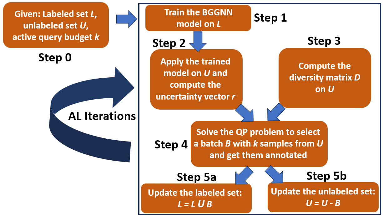 Empowering Active Learning for 3D Molecular Graphs with Geometric Graph IsomorphismAdvances in Neural Information Processing Systems, 2024
 -
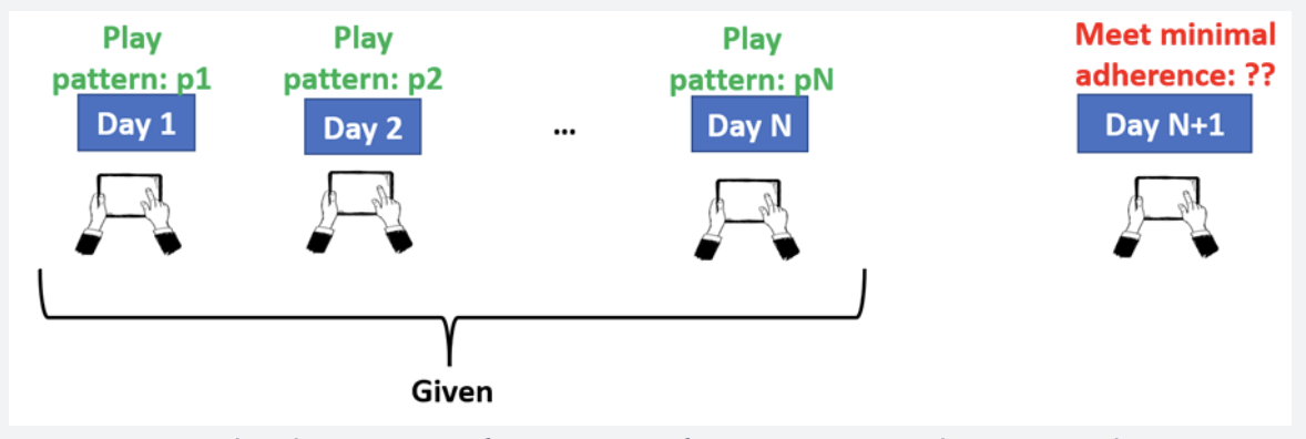 Predicting Adherence to Computer-Based Cognitive Training Programs Among Older Adults: Study of Domain Adaptation and Deep LearningJMIR aging, 2024


